Author Archive

Stroke Rehabilitation: Importance, Methods, & Cardiologist Insights

Stroke Rehabilitation is VITAL after. It should be minimum 15 hours a week with 7 day working

Why is a cardiologist interested in stroke?

Firstly because it is a vascular disease.

Secondly because there is a risk of stroke with the procedures I do. There is no point is a perfect heart and then a stroke that debilitates my patient.

Thirdly, I want the best treatment for my patient. I want acute stroke intervention if they have had a stroke (in my mind that is Mechanical Thrombectomy), and I want the best stroke rehabilitation so they get the best recovery.

I have had a stroke- should I join a rehabilitation program?

The short answer is yes. Just like for cardiac rehabilitation after stroke (which reduces your risk of death by 17% compared to not having it), stroke rehabilitation is evidence based.

It needs to be planned BEFORE you go home. It should cover YOUR needs. That may be to do with movement, speech, hearing, or fatigue. It could also involve memory.

Why am I so fatigued after my stroke?

This is very common after stroke and under recognised. In most it takes a year to get better. In 40% it can take 2 years. It can be the ONLY and WORST symptoms you have. It is separate from low mood or depression.

Vision and hearing issues

This may not be obvious and needs formal assessment. It needs to involve your family also. They may have noticed a subtle change that needs to be addressed.

What about community participation programs?

Quality of life is improved in general by these- which include physical exercise, art, music. Whatever it is, it needs to be what YOU need. There is good evidence for both group exercise and individual programs when it comes to physical therapy.

How much and how often?

Motor therapy of 1-2 hours per day started EARLY, and sustained, improved recover in 1st 6 months. Delayed initiation has less benefit but still helps. Better later than never!

Physiotherapy, Occupational and speech and language therapy (SALT) should be at least 3 hours a day and at least 5 days a week. Less is not as good. So aim for 15 hours a week or more.

Where can I find out more?

This is a summary of the recent NICE guidance. See the full guidance here.


Book your consultation

f you are worried about a stroke or your heart and are struggling to decide what to do, please book a consultation, I will be more than happy to see you. Let’s get the quality of your life back.

Article by Dr Malik, a UK leading cardiologist. He works at One Welbeck Heart Health – London’s Largest Private Cardiology Group, and at Hammersmith Hospital, Imperial College Healthcare NHS Trust, London, one of the largest NHS Trusts in the UK.

 

Does the PREVENT trial really show stents can prevent heart attacks?

The short answer is maybe, but if it was my heart, I am not yet rushing to have more stents put in.

Should I get a stent in a coronary artery that is not significantly narrow?

Firstly, what is a significant narrowing? Anatomically it can look “tight” and if it looks 90% narrow it probably is. But if it looks 50-80% narrow, physiological testing will be needed to confirm it is indeed flow limiting.

Secondly, just because it is narrow, it does not mean a stent is needed. Except for the left main stem artery (LMS) and left anterior defending artery (LAD), there is no evidence that stenting will make you live longer.

Unless you have terrible angina, it won’t make you feel better either!

What is a “vulnerable plaque”?

Why do some plaques drive to rupture and block the blood vessels? They are more inflamed and more unstable.  They can’t be seen from outside the body. There is data that they can be images from inside the blood vessel.

We can’t see the inflammation site you but can estimate it.


Thus:

  • Thin cap on top of it- so it is more fragile
  • A high content of fat in the plaque – so it is more fragile
  • A higher burden of plaque- there is more of it in that spot- more than 70% burden
  • A narrower vessel – there is more of it and it needs to increase in size less to block off the blood vessel – less than 4mm2 area minimum

This can be assessed with intravascular ultrasound (IVUS) or with optical coherence tomography (OCT). This itself requires skill in doing it and interpreting it.  I do imaging routinely when performing stenting, but these investigators also looked at areas >50% narrowed on angiogram, but with no flow limitation (measured with FFR >0.8).

They used the above 4 criteria. If two were present, then it was a vulnerable plaque.

Interestingly half of patients with over 50% narrowing with FFR >0.8 had “vulnerable plaques”.

What does this trial say?

Over a 2 year follow-up, 1606 patients were randomised and assessed for the primary endpoint of: a composite of death from cardiac causes, target-vessel myocardial infarction, ischaemia-driven target-vessel revascularization, or hospitalisation for unstable or progressive angina,

That means that any of these could have led to a difference.

At 2 years:

Primary outcome :
-3 (0·4%)  in the stent group versus 27 (3·4%)  in the medical therapy group (absolute difference –3·0 percentage points [95% CI –4·4 to –1·8]; p=0·0003). This means it was highly statistically significant

Death:
-4 (0·5%) versus 10 (1·3%) patients (absolute difference –0·8 percentage points [95% CI –1·7 to 0·2]). This was NOT Statistically significant.

Heart attack: 
-nine (1·1%) versus 13 (1·7%) patients had myocardial infarction (absolute difference –0·5 percentage points [–1·7 to 0·6]). This was NOT Statistically significant.

Issues with the trial:

The largest issue is that it was not blinded – thus the patient and the doctor knew in the non-interventional arm that there was a “vulnerable plaque”- that makes THEM vulnerable to the suggestion that there was a risk, and thus any “angina” should be taken seriously.

Secondly, it did not prove that death or heart attacks were significantly reduced – those are hard endpoints that cannot be affected by bias.

Thirdly, patients with stents were given strong anti-platelet medication that could have reduced risk. And lastly, 50 patients (3%) were lost to follow-up. This may have affected the results

So, it is interesting- but will it make me change my view today- no. I will NOT be stenting vulnerable plaques tomorrow.


How do I find out more?:

You can read the paper on the study here.

Book your consultation

If you have asymptomatic Coronary Artery Disease and are struggling to decide what to do, please book a consultation, I will be more than happy to see you. Let’s get the quality of your life back.

Article by Dr Malik, a UK leading cardiologist. He works at One Welbeck Heart Health – London’s Largest Private Cardiology Group, and at Hammersmith Hospital, Imperial College Healthcare NHS Trust, London, one of the largest NHS Trusts in the UK.

Asymptomatic Coronary Artery Disease | What Should You Do?

Asymptomatic Coronary Artery Disease Explained

So, you’ve got asymptomatic coronary artery disease. Should you do something or nothing? Dr Malik runs through your options, breaking down the complexities in a simple way.

What is Coronary Artery Disease (CAD)?

Coronary Artery Disease (CAD) is a condition involving the coronary arteries. You have a left and right coronary artery. These are the first branches that come off the aorta, and they supply blood to your heart. Normally, they have smooth internal linings and can expand to increase blood supply during physical activities, like exercise. If you start having atherosclerosis (furring up), they can become narrow, meaning they fail to supply enough blood to the heart during exercise. 

Are there any symptoms I should be watching out for?

Angina is the most common symptom of CAD. It’s the feeling of “pain”, discomfort, breathlessness or tightness that is brought on with exercise and is a result of the heart not getting enough blood. In some patients, the heart can feel the strain, but you may not feel the pain! This is because pain is a subjective feeling. But also, the symptoms can be confused with indigestion, heartburn, being unfit, getting older… or of course, you may truly have no symptoms.

How is asymptomatic CAD screened for?

We can use several methods to screen for CAD.

  • Stress test: to check for any strain on your heart.
  • CT coronary angiogram: to check for any evidence of furring up – this is often the way we pick up more minor narrowings that may not cause symptoms
  • Invasive angiogram: to diagnose CAD and measure the disease’s severity

Coronary Artery Disease treatment options

If you do have Coronary Artery Disease, there are several treatment options.

1. Medical therapy only

  • Anti-platelets such as aspirin. There’s good evidence in younger patients, but more side effects in older patients.
  • Statins to lower lipids. Statins carry a lower risk and are worth it – I am on a statin!
  • Anti-anginal pills (but only if you have symptoms of angina):
    • Nitrates
    • Beta-blockers
    • Calcium channel blockers
    • Nicorandil
    • Ranolazine
    • Colchicine – an anti-inflammatory that’s now useful in treating coronary disease

2. Combining medical therapy with angioplasty & stenting

Medical therapy plus angioplasty and stenting (also known as PTCA or PCI is a less invasive option).  Angioplasty is usually done through the wrist and involves placing a stent or scaffold to open up the blockage. The latest data shows that historically, we’ve carried out too much angioplasty, so please be aware that angioplasty is only really for symptom control. Outside of having an acute heart attack, stents are unlikely to extend your lifespan.

3. Combining medical therapy with coronary bypass surgery (CABG)

If you have severe CAD, Coronary Bypass Surgery offers a “full replumbing” and may help you live longer. 

There are different types of surgeries: 

  • On pump surgery vs off pump surgery
  • Mini access surgery vs open chest surgery
  • Full arterial revascularisation vs using a LIMA graft and veins

But you shouldn’t worry too much about the options – our private heart specialists are here to advise on the right treatment that improves your quality of life. 

How do we decide which treatment is best?

We consider the following questions:

  1. Are you robust/strong enough to take the treatment offered? 
  2. What does the evidence from trials, medical therapy and PCI versus CABG show? You may only need to improve your lifestyle, weight and blood pressure. 
  3. What do YOU want? You may want a less invasive treatment, but if that’s significantly worse for you, we may choose not to offer that to you. Our cardiologists only advise on procedures that are going to be best for you and your quality of life.

Who finally chooses?

You’re allowed to make a bad choice – it’s your body after all. But we don’t have to carry out a certain treatment if we disagree. You may find another cardiologist who’ll do it, but that won’t change the evidence base or facts.  Real patients are harder to manage than trial patients. The Multidisciplinary team (MDT) is useful when a plan is not clear-cut – but in the end, you and your own doctor have to agree on a plan!


Watch Dr Malik delivering a talk to Cardiothoracic surgical trainees and running through the evidence – should you do something, or nothing? 

Book your consultation

If you have asymptomatic Coronary Artery Disease and are struggling to decide what to do, please book a consultation, I will be more than happy to see you. Let’s get the quality of your life back.

Article by Dr Malik, a UK leading cardiologist. He works at One Welbeck Heart Health – London’s Largest Private Cardiology Group, and at Hammersmith Hospital, Imperial College Healthcare NHS Trust, London, one of the largest NHS Trusts in the UK.
Paravalvular Leak | A Challenging Case With Dr Malik

How Dr Malik Closed A Challenging Paravalvular Leak

Dr Malik talks through a challenging case, explaining what a paravalvular leak is and the best available treatments. 

With one of the UK’s largest experiences of closing paravalvular and other odd leaks, he’s got the expertise to keep your mind at ease and help you avoid the need for surgery. Let’s delve into the case study.

Watch Dr Malik discuss a tricky case of Paravalvular leak

Video: Click Here

“Watch me deliver a presentation to a group of surgeons on this tricky case. A complication of open surgical valve replacement, with a tricky hole was left behind. I managed to plug it and reduce the leak. The patient has felt so much better ever since!”

What is a paravalvular leak?

After surgical valve replacement, leaks can sometimes occur. Either THROUGH  the valve – or  BY THE SIDE  of it – due to a weakness in the tissues which allows the sutures to loosen. If the leak is severe enough, the valve can even start “rocking”. 

In this particular case that I presented, however, a suture seemed to have created an additional hole, connecting the left ventricle to the right atrium. This is what’s known as a Gerbode Defect.

What treatments are there for Paravalvular Leak?

There are several options:

  • Medical therapy. If you’ve got no symptoms and the heart is stable, it may not be necessary to close the leak. Not every hole needs closing.
  • Transcatheter device placement. Plug it with a transcatheter device put in from the leg.
  • Redo surgery. If the surgical risk isn’t too high and a plug’s not possible, re-doing the surgery will likely be your best option.

What tests do I need?

For a suspected paravalvular leak, you’ll need a full assessment. This includes:

Worried about your heart?

Please book a consultation or contact us at DrMalikPA@OneWelbeck.com. With our patient-centred approach, you’ll feel confident you’re in safe hands.

Article by Dr Malik, a UK leading cardiologist. He works at One Welbeck Heart Health – London’s Largest Private Cardiology Group, and at Hammersmith Hospital, Imperial College Healthcare NHS Trust, London, one of the largest NHS Trusts in the UK.
What is the Mitral Valve? | Dr Malik Explains

Mitral Valves Explained by UK Leading Cardiologist

Learn all about Mitral Valves with Dr Malik – one of the UK’s leading heart surgeons, known for his simple explanations of complex heart problems. Let’s explore what the mitral valve is, which side it’s located and the best treatment options for when it leaks.

What is a Mitral valve?

The Mitral valve is the inflow valve of the heart, located on the left-hand side. It has two leaflets that open and close in sync with the heartbeat.  Mitral valves can encounter issues such as narrowing, typically caused by Rheumatic Heart disease. It can also experience leakage, often due to problems at the valve’s base that cause it to lose support, or issues with the leaflets not meeting properly (leading to the blood leaking backwards).

What’s the best treatment for problems with the mitral valve?

The treatment options depend on the problem.

Stenosis (or narrowing)

A balloon can be used in younger patients to open it up. In older patients, surgery is usually needed.

Regurgitation or leaking

Regurgitation or leaking in the Mitral valve can be treated with surgery, typically an open valve repair or replacement. But if surgery is high-risk, a “clip” can be placed on the valve to reduce (but not cure) the leak.

Issues with a failing surgical valve

When a surgical valve begins to fail, there are a few potential issues we must consider.

  • Leakage around the valve’s edge. If there’s a leakage around the valve’s edge, one option is to plug the leak from the leg. Alternatively, another round of surgery may fix the issue.
  • Narrowing or leaking inside the valve.  The treatment for internal valve leaking depends on the type of valve.
  • Metallic valve. Another round of surgery will almost always be required.
  • Tissue valve. A TAVI valve could be inserted. Dr Malik is a national proctor for this procedure and teaches others how to do it.

How do we assess the mitral valve?

The mitral valve can be listened to with a stethoscope. It makes sounds (murmurs) if it’s NOT working well. If we suspect a problem, we’ll order more tests, such as:

 Watch Dr Malik carry out a malfunctioning Mitral valve replacement

Dr Malik carries out these procedures routinely. Watch and learn:

Click Here

“I performed this procedure under general anaesthetic via a small puncture in the groin. The latest technology can achieve outstanding results, even in high-risk patients!”

Worried about your heart?

Please book a consultation or contact DrMalikPA@OneWelbeck.com to put your mind at ease and get the latest, high-quality tests your heart deserves.

Article by Dr Malik, a UK leading cardiologist. He works at One Welbeck Heart Health – London’s Largest Private Cardiology Group, and at Hammersmith Hospital, Imperial College Healthcare NHS Trust, London, one of the largest NHS Trusts in the UK

How We Helped Amy Recover From Her Stroke Back To Full Health

Learn how Dr Malik helped Amy recover from her stroke and go back to full health. She’s now happy, healthy and fully active! 


Amy’s had a very good recovery and kindly agreed to have the procedure filmed.

Watch her journey below.


Amy Jones is an Instagram influencer who had a PFO closure at One Welbeck Heart Health with Dr Iqbal Malik – who leads the NHS Structural Heart Service at Imperial College Healthcare NHS Trust.

Dr Malik says: “We decided to close the PFO, and Amy agreed for it to be filmed to help other people who were due to have the procedure. We used a Cocoon device. It went well and Amy is now back to full health and fully active! I am very grateful for her help in making this video”

Investigations

First, Amy had a thorough series of investigations to diagnose the cause of her stroke.

What were the results?

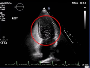
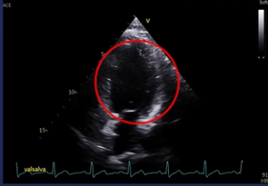
The Bubble Echocardiogram showed she had shunting of the microbubbles of the right side of the heart across to the left side. Even spontaneously at rest. Amy’s stroke appeared to be caused by the PFO (Patent Foramen Ovale) – a flap valve in the heart that is also present in 25% of healthy people. This meant Amy was suitable for a PFO Closure to significantly lower the risk of another stroke.

A straightforward procedure

The team made Amy feel comfortable and relaxed throughout the investigations and procedure.

When she woke up, Dr Malik told her: “The procedure went really well. As straightforward as a PFO closure can go really! The tunnel was really quite small, so I’m hopeful that in 2 weeks’ time, the button will have really sealed up and the bubble study will be negative. Then we can forget about the PFO and get on with life.”

Amazing aftercare

When Amy got home from her procedure, the friendly specialist nurse Helda gave her a call to make sure everything was okay. Amy told her she had a bit of pain, and Helda reassured her that soreness in the throat and groin are expected, as well as some minor palpitations. This made Amy feel very at ease knowing her recovery is on the right track. We offer full support after you leave the clinic, which our patients really value.

Back to full health

Fortunately, Amy did really well and only 2 weeks after the procedure, we confirmed with a repeat bubble echocardiogram that she was fully fixed!

BEFORE

Before a PFO

AFTER

After a PFO

2 weeks after the procedure, there are no longer any bubbles: at rest, with a sniff, or even with a strong Valsalva (straining). Amy is now happy, healthy and fully active without having to worry about her PFO.

Have you got a PFO?

If you have a PFO and want Dr Malik’s help, visit our Contact Us page and make an enquiry today. Join Amy and get the quality of your life back!